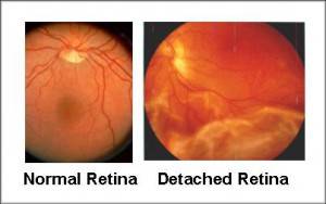

Treatment usually prevents retinal detachment. These treatments cause little or no discomfort and may be performed in your ophthalmologist’s office.

Most retinal tears need to be treated with laser surgery or cryotherapy (freezing), which seals the retina to the back wall of the eye. Only after careful examination can your ophthalmologist tell whether a retinal tear or early retinal detachment is present. Some retinal detachments are found during a routine eye examination. Your ophthalmologist can diagnose retinal detachment during an eye examination in which he or she dilates (enlarges) the pupils of your eyes. These symptoms do not always mean a retinal detachment is present however, you should see your ophthalmologist as soon as possible. a gray curtain moving across your field of vision.a shadow in the periphery of your field of vision.These early symptoms may indicate the presence of a retinal detachment: What are the warning symptoms of retinal detachment? weak areas in your retina that can be seen by your ophthalmologist (Eye M.D.).previous retinal detachment in your other eye.The following conditions increase the chance of having a retinal detachment: Fluid may pass through the retinal tear, lifting the retina off the back of the eye, much as wallpaper can peel off a wall. But sometimes the vitreous pulls hard enough to tear the retina in one or more places. Usually the vitreous separates from the retina without causing problems. As we get older, the vitreous may pull away from its attachment to the retina at the back of the eye. A clear gel called vitreous (vit-ree-us) fills the middle of the eye.


 0 kommentar(er)
